Introduction
Ultrasound-guided sclerotherapy (UGS) is a highly specialized procedure for treating varicose veins that are hidden beneath the skin.
Not all varicose veins are visible and some feed directly into the spider veins on the surface of the skin. In these cases, ultrasound is used to identify the target vein, and the medication is injected into the desired segment. Vein doctors adept at ultrasound techniques are best suited to perform this advanced technique for vein removal.
How Does Ultrasound Guided Sclerotherapy Work?
A detailed ultrasound examination is performed prior to sclerotherapy to create a road map of the lower leg veins. This map precisely displays all abnormal veins and helps the vein doctor identify the root cause of the visible spider veins and varicose veins.
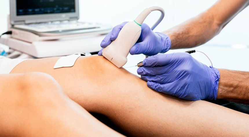
Using ultrasound guidance, the doctor can accurately guide the needle and inject the sub-surface veins underlying visible ones. The ultrasound also helps identify regions of deep venous connections (perforator veins) that are carefully monitored during treatment. Once injected, the treated vessel will collapse and gradually be absorbed by the body, disappearing with time.
Foam sclerotherapy is well visualized on ultrasound and allows for real-time monitoring of treatment. The foam is better suited for larger varicose veins than liquid sclerotherapy as it maintains better contact with the vessel walls. Additional safety enhancements include pre-formulated compounds with O2:CO2 mixtures that micronize bubbles leading to more uniform treatment. In our vein clinic, we routinely perform ultrasound-guided sclerotherapy for large visible varicose veins, neovascularization, and residual segments of saphenous veins that are tortuous or in close proximity to vulnerable structures like skin and nerves. It is used in conjunction with vein ablation for the best results.
What Happens During Ultrasound Guided Sclerotherapy?
- The target vein segments are mapped with ultrasound. Entry sites and perforator veins are marked on the skin for special attention.
- The target vein(s) is accessed using ultrasound guidance, and the sclerosant is injected into the vein
- Foam sclerotherapy is visualized in real-time to monitor treatment response. Additional injections are given based on this information. The leg is often elevated during the injection to increase the treatment response
- Smaller spider veins may be treated during the same session or during follow-up for cosmetic sclerotherapy